Back Muscles Anatomy Reference / Intrinsic Back Muscles Anatomy Of The Torso Medical Library / Jun 29, 2016 · the breast lies over the musculature that encases the chest wall.
Back Muscles Anatomy Reference / Intrinsic Back Muscles Anatomy Of The Torso Medical Library / Jun 29, 2016 · the breast lies over the musculature that encases the chest wall.. The blood supply that provides circulation to these muscles perforates through to the breast parenchyma, thus also supplying blood to the breast. Tightening (contraction) or relaxing these muscles causes the lens to change shape, allowing the eyes to focus on near or far objects (accommodation). Techniques edit May 27, 2015 · the referral pattern of the rhomboids is not as widely distributed but local to the muscles. May 31, 2021 · try out our muscle anatomy reference charts with the attachments, innervation and actions of all 600+ muscles of the human body, all in one place.
The shoulder is a complex joint with high mobility but a lack of stability. May 31, 2021 · try out our muscle anatomy reference charts with the attachments, innervation and actions of all 600+ muscles of the human body, all in one place. One particular feature of the digastric is that both bellies have different embryological origins. Techniques edit The muscles involved include the pectoralis major, serratus anterior, external oblique, and rectus abdominis fascia.
Anatomy Of The Back Spine And Back Muscles Kenhub from thumbor.kenhub.com This group of muscles comprise the semispinalis, multifidus, and rotatores muscles. Small muscles attached to the lens can change its shape, allowing the eye to focus on objects at varying distances. The vitreous chamber is between the lens and the back of the eye. One particular feature of the digastric is that both bellies have different embryological origins. The shoulder is a complex joint with high mobility but a lack of stability. The anatomical planes are different lines used to divide the human body. Both work together to produce normal movements. Here we explain the basic anatomy of the shoulder.
One particular feature of the digastric is that both bellies have different embryological origins.
Both work together to produce normal movements. The anatomy of the shoulder consists of the shoulder joint and shoulder girdle. Flexes the metacarpophalangeal, extends proximal and distal interphalangeal joints and adducts digits 1, 2, 4, & 5 (adduction of the digits of the hand is in reference to the midline of the 3rd digit) ulnar nerve, deep branch : The pain generally extends from the edge of the shoulder blades to the spine. Small muscles attached to the lens can change its shape, allowing the eye to focus on objects at varying distances. Techniques edit Tightening (contraction) or relaxing these muscles causes the lens to change shape, allowing the eyes to focus on near or far objects (accommodation). A plane is a 2d slice through 3d space, which can be thought of as a glass sheet. The blood supply that provides circulation to these muscles perforates through to the breast parenchyma, thus also supplying blood to the breast. May 31, 2021 · try out our muscle anatomy reference charts with the attachments, innervation and actions of all 600+ muscles of the human body, all in one place. The muscles involved include the pectoralis major, serratus anterior, external oblique, and rectus abdominis fascia. The vitreous chamber is between the lens and the back of the eye. Jun 29, 2016 · the breast lies over the musculature that encases the chest wall.
May 31, 2021 · try out our muscle anatomy reference charts with the attachments, innervation and actions of all 600+ muscles of the human body, all in one place. The anatomical planes are different lines used to divide the human body. The blood supply that provides circulation to these muscles perforates through to the breast parenchyma, thus also supplying blood to the breast. Here we explain the basic anatomy of the shoulder. A plane is a 2d slice through 3d space, which can be thought of as a glass sheet.
Back Muscles In A Nutshell 3d Models Video Tutorials Notes Anatomyzone from anatomyzone.com The pain generally extends from the edge of the shoulder blades to the spine. Jun 29, 2016 · the breast lies over the musculature that encases the chest wall. The anatomical planes are different lines used to divide the human body. The anatomy of the shoulder consists of the shoulder joint and shoulder girdle. Here we explain the basic anatomy of the shoulder. The blood supply that provides circulation to these muscles perforates through to the breast parenchyma, thus also supplying blood to the breast. The shoulder is a complex joint with high mobility but a lack of stability. Techniques edit
Small muscles attached to the lens can change its shape, allowing the eye to focus on objects at varying distances.
Jun 29, 2016 · the breast lies over the musculature that encases the chest wall. All of these muscles are subdivided further according to the region they span. Small muscles attached to the lens can change its shape, allowing the eye to focus on objects at varying distances. Techniques edit The anatomical planes are different lines used to divide the human body. The blood supply that provides circulation to these muscles perforates through to the breast parenchyma, thus also supplying blood to the breast. Tightening (contraction) or relaxing these muscles causes the lens to change shape, allowing the eyes to focus on near or far objects (accommodation). May 31, 2021 · try out our muscle anatomy reference charts with the attachments, innervation and actions of all 600+ muscles of the human body, all in one place. Here we explain the basic anatomy of the shoulder. A plane is a 2d slice through 3d space, which can be thought of as a glass sheet. The vitreous chamber is between the lens and the back of the eye. This group of muscles comprise the semispinalis, multifidus, and rotatores muscles. The muscles involved include the pectoralis major, serratus anterior, external oblique, and rectus abdominis fascia.
Small muscles attached to the lens can change its shape, allowing the eye to focus on objects at varying distances. The anatomical planes are different lines used to divide the human body. Jun 29, 2016 · the breast lies over the musculature that encases the chest wall. The pain generally extends from the edge of the shoulder blades to the spine. This group of muscles comprise the semispinalis, multifidus, and rotatores muscles.
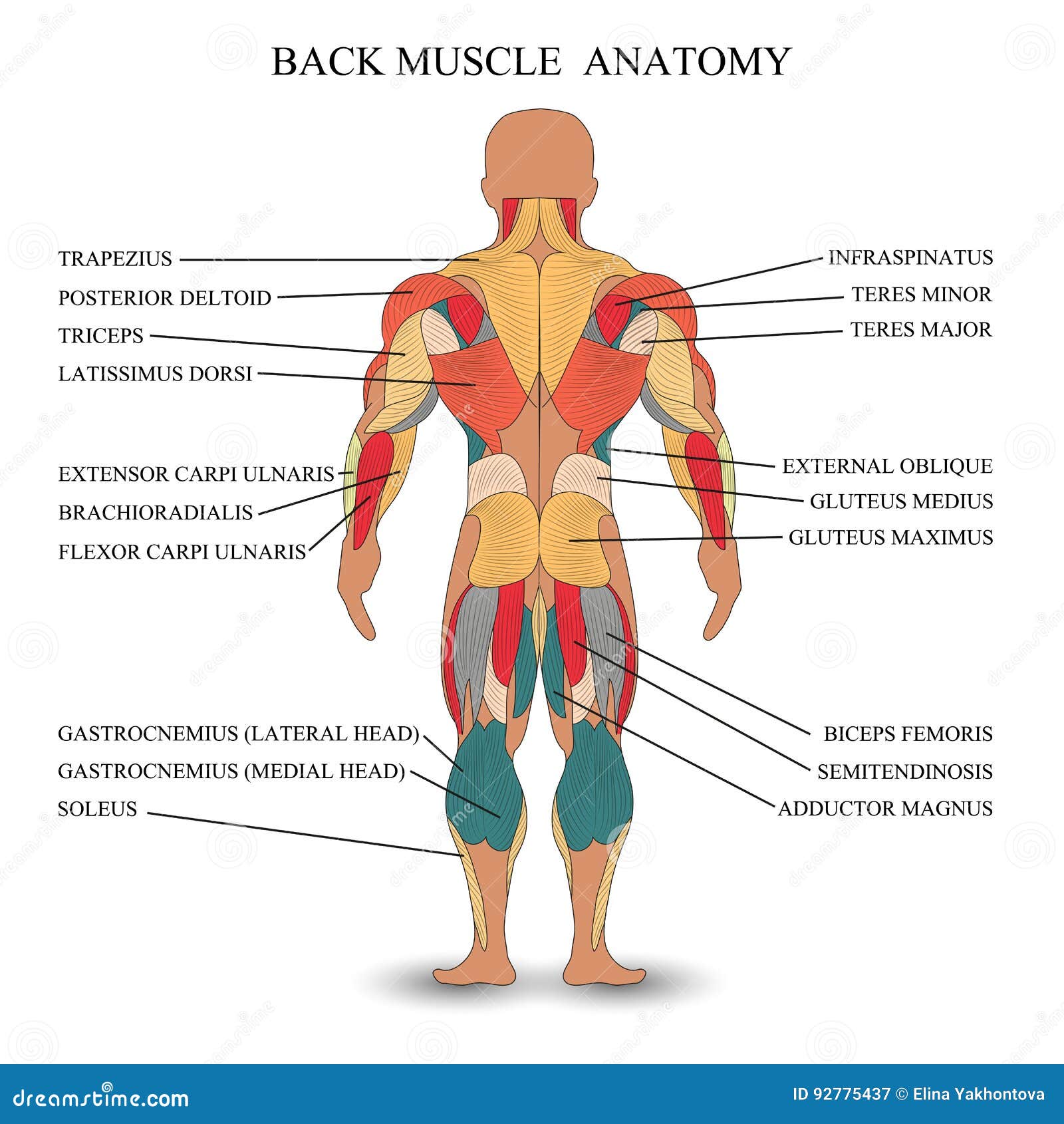
Anatomy Back Muscles Stock Illustrations 1 418 Anatomy Back Muscles Stock Illustrations Vectors Clipart Dreamstime from thumbs.dreamstime.com Jun 29, 2016 · the breast lies over the musculature that encases the chest wall. All of these muscles are subdivided further according to the region they span. The blood supply that provides circulation to these muscles perforates through to the breast parenchyma, thus also supplying blood to the breast. Flexes the metacarpophalangeal, extends proximal and distal interphalangeal joints and adducts digits 1, 2, 4, & 5 (adduction of the digits of the hand is in reference to the midline of the 3rd digit) ulnar nerve, deep branch : The anatomical planes are different lines used to divide the human body. A plane is a 2d slice through 3d space, which can be thought of as a glass sheet. Both work together to produce normal movements. May 31, 2021 · try out our muscle anatomy reference charts with the attachments, innervation and actions of all 600+ muscles of the human body, all in one place.
May 27, 2015 · the referral pattern of the rhomboids is not as widely distributed but local to the muscles.
Techniques edit May 27, 2015 · the referral pattern of the rhomboids is not as widely distributed but local to the muscles. The anatomical planes are different lines used to divide the human body. Since the rhomboids on both sides are almost always affected, this is a primary source of mid back tightness or aching between the shoulder blades. The shoulder is a complex joint with high mobility but a lack of stability. The anatomy of the shoulder consists of the shoulder joint and shoulder girdle. Tightening (contraction) or relaxing these muscles causes the lens to change shape, allowing the eyes to focus on near or far objects (accommodation). Here we explain the basic anatomy of the shoulder. Flexes the metacarpophalangeal, extends proximal and distal interphalangeal joints and adducts digits 1, 2, 4, & 5 (adduction of the digits of the hand is in reference to the midline of the 3rd digit) ulnar nerve, deep branch : All of these muscles are subdivided further according to the region they span. A plane is a 2d slice through 3d space, which can be thought of as a glass sheet. Jul 26, 2021 · the transversospinalis muscles are a large group of muscles that also belong to the deep layer of the intrinsic muscles of the back. Small muscles attached to the lens can change its shape, allowing the eye to focus on objects at varying distances.


0 Comments